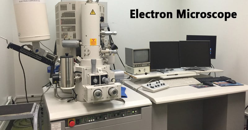
Scanning and transmission electron microscopy is a very significant tool in scientific & engineering societies. It is particularly useful when high magnification inspections have to be done. The scanning electron microscope resolution not only allows for very high magnification but also offers outstanding depth of field. And, this high magnification-depth of field combination is particularly useful in the field of failure analysis where both of the aspects have great importance. When examining the fracture surfaces in the attempt to identify failure mode, investigating engineers requires having a clear view of fracture features, oftentimes at very high magnifications. Features such as fatigue striations are very small & need magnifications in excess of 1000X for clear identification. Various other unexpected constituents like un-wanted inclusions & other defects can often time are eagerly observed with high magnifications offered by the scanning electron microscope.
Along with real-time aspects involved with the scanning electron microscope operation, documentation is very significant in communicating failure analysis results with the customers, attorneys, etc. The modern scanning electron microscope allows for digital photographic documentation that can include specific labeling & even the measurement of significant geometric structures. In addition, many if not most SEM’s are furnished with the capability to perform Energy Dispersive Spectroscopy chemical analysis. Areas or even precise particles of interest can be analyzed by using the Energy Dispersive Spectroscopy system while the sample is being examined at high magnification. An Energy Dispersive Spectroscopy spectrum can be generated that indicates the general chemical make-up of the area or particle of interest. The Energy Dispersive Spectroscopy spectrum can also be used in the final failure analysis report when the chemical composition is of importance.
Usually, all of the information & data collected during scanning electron microscopy examination by environmental scanning electron microscope is combined with other evaluations performed during the failure analysis & is presented in the detailed formal test report. The other information collected may include metallurgical test results, visual & stereo microscope findings, hardness measurements, and any history regarding the service conditions prior to or during the failure. This is not uncommon for the investigating engineer to re-examine certain aspects of physical evidence as the conclusions become evident. The data & information included in the final summary should all lead to the final conclusion regarding failure mode. The scanning electron microscope is the best method for examining fracture surfaces at high magnification. It will continue to be a more & more important tool in performing the failure analysis as its value continues to be recognized & as the number and capabilities of scanning electron microscopes continue to grow. In the scanning electron microscope, the image was made based on the detection of new secondary electrons, or reflected electrons emerging from the sample surface when the sample surface was scanned by the electron beam.
Benefits of the Scanning Electron Microscope
Research is becoming more & more advanced on a daily basis, and these kinds of magnifying instruments are certainly needed, no matter what the cost. The advantages severely outweigh any disadvantages which one might think of when it comes to owning a scanning electron microscope. The top advantage of a scanning electron microscope is the depth of field that the device offers. By being able to study the entire object instead of just part of the item, one can learn more from the microscope images. The scanning microscope is one of the few microscopes in the market which offers this type of depth. This microscope also needs less sample preparation than other types, like a transmission electron microscope. This can save time which can go to focusing on study, rather than prep of a specimen.
Numerous advantages exist when choosing the scanning microscope for the laboratory. The microscope provides extremely high resolution of images & this is vital in today’s world of research. This also provides a higher level of magnification than most other microscopes & this helps to ensure images are seen in minute detail. This can help in studying numerous viruses & even parts of an animal or insect. Since the microscope uses electromagnets instead of the lenses, the user has an uncanny ability to see items at a much closer range. This can prove vital when studying and trying to explore cures for diseases. The scanning electron microscope can mean the difference between a laboratory & a first-rate one. If the scientist wants to be on the cutting edge of technology in the scientific community, then the scanning microscope can prove to be invaluable.
How much is an electron microscope cost? Even though they are very expensive, a good laboratory cannot be without one. In order to ensure that this is the appropriate choice for one & their needs, a person should learn all they can about all such great magnifying instruments. Even though a scanning electron microscope is not for everyone, for those who study to cure, this type is definitely a must-have in the laboratory.


_3-6.jpg)


